The Legacy of Bolognese Wax Art: Giuseppe Astorri and Cesare Bettini
Giuseppe Astorri
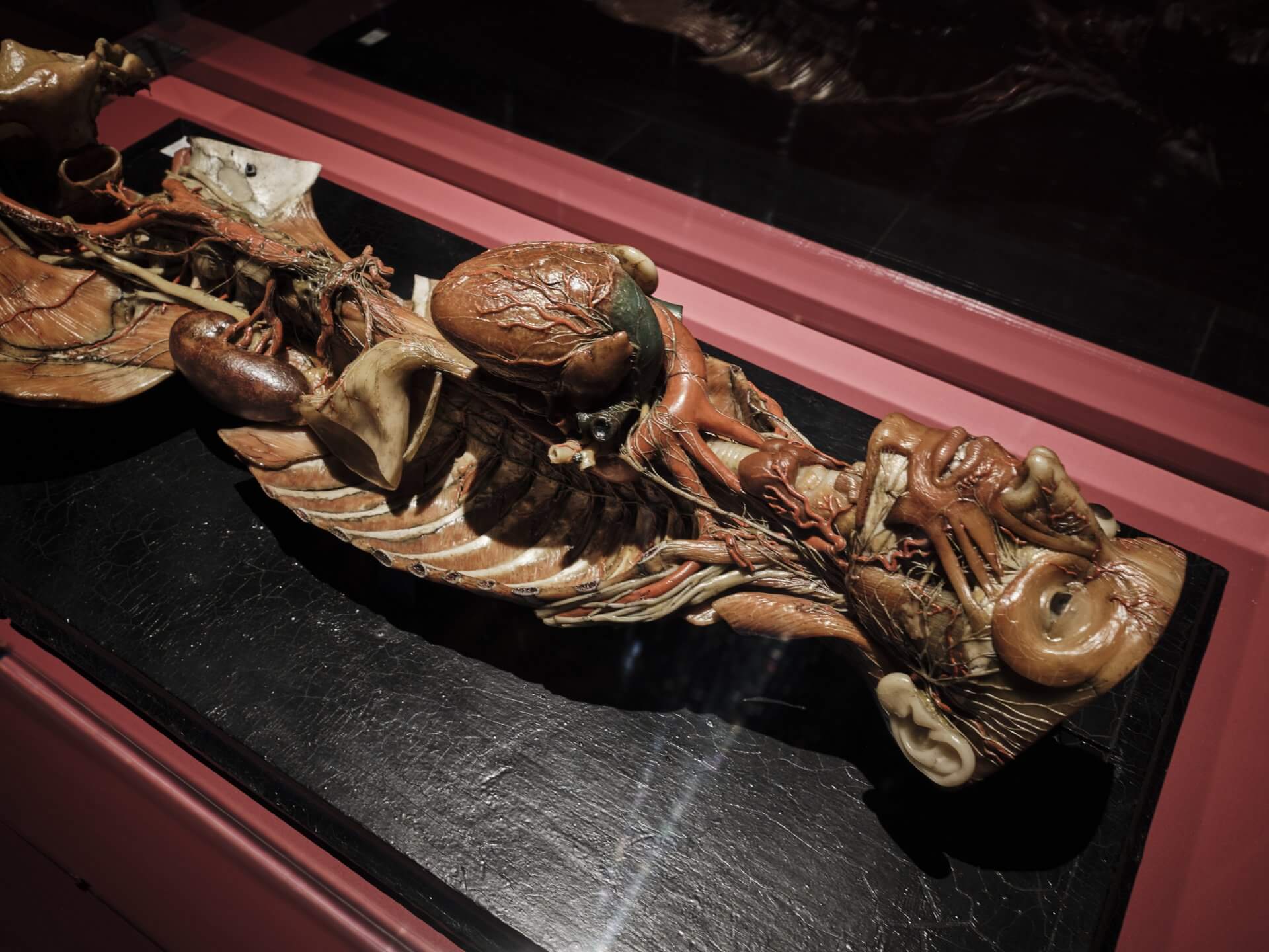
Testa e tronco di donna con dimostrazione del circolo arterioso e del sistema simpatico (Head and torso of a woman with a demonstration of the arterial circulation and the sympathetic nervous system), 18th–19th c.
Anatomical model in wax painted using mixed media
With frame 57,5 x 39 x 20 cm
Cesare Bettini
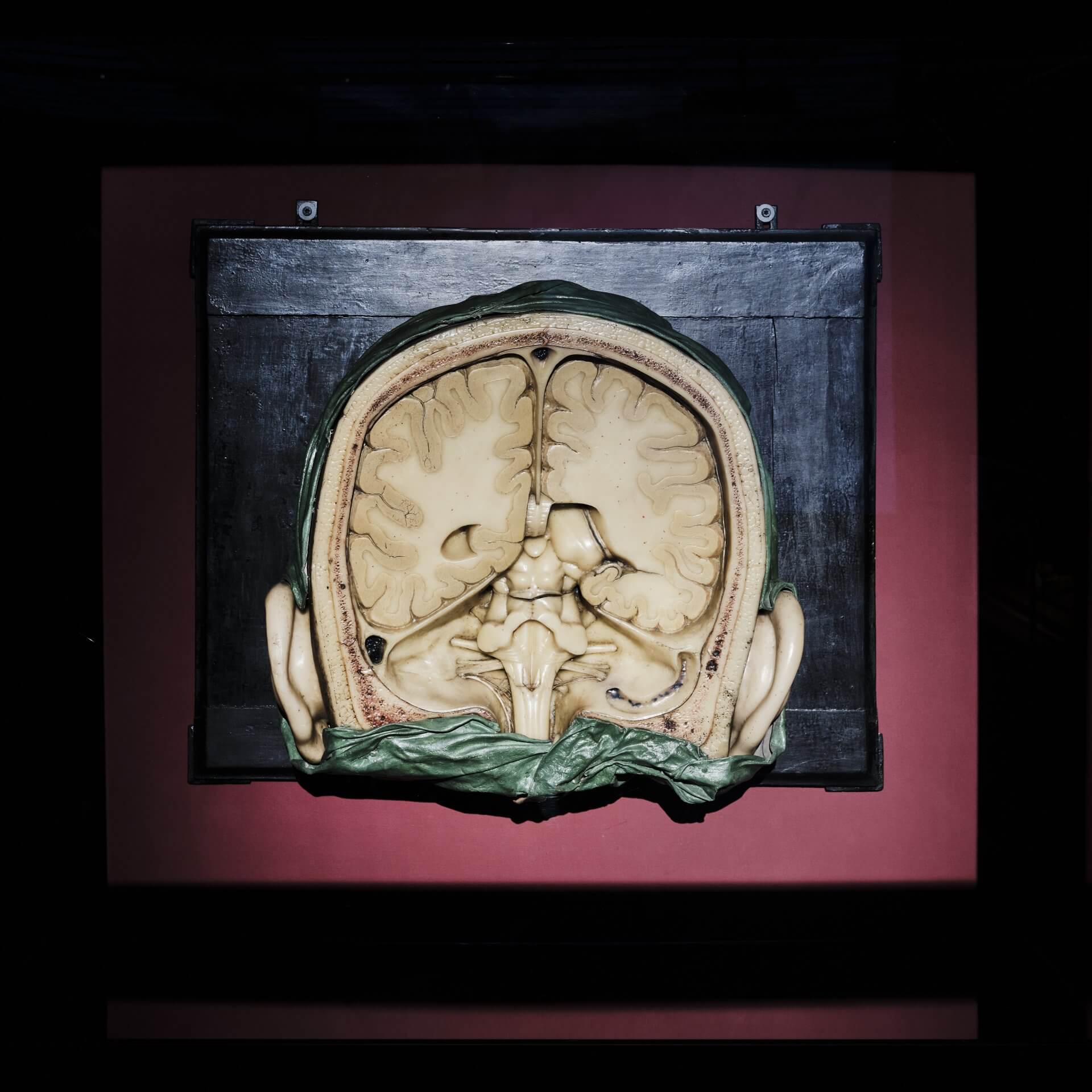
Sezione frontale del cranio con gli emisferi cerebrali sezionati e del tronco encefalico visti
posteriormente (Frontal section of the skull with the cerebral hemispheres dissected and the brainstem seen from behind)
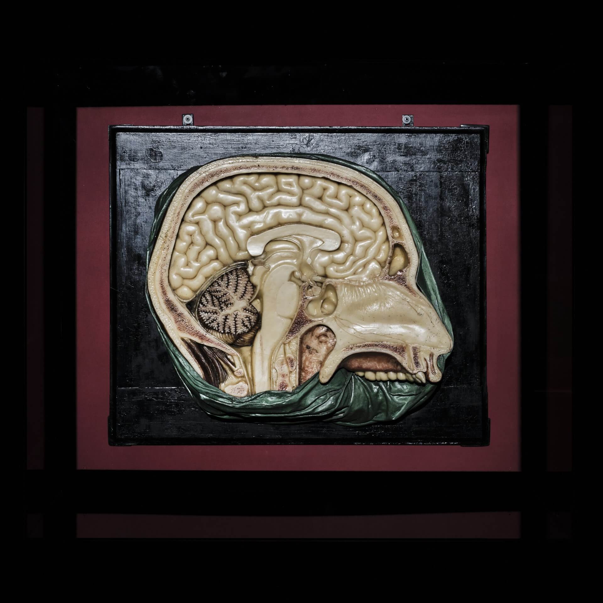
Modello di sezione sagittale del cranio mostrante le parti dell’encefalo (Model of a sagittal section of the skull showing the parts of the brain), 19th c.
Two Anatomical models made of painted wax, wood, fabric
With frames 88 x 72 x 34 cm; 101 x 85 x 26 cm
Courtesy Alma Mater Studiorum – University of Bologna | University Museum Network | “Luigi Cattaneo” Anatomical Wax Collection
From the “Luigi Cattaneo” Collection of the University of Bologna come three wax models by Giuseppe Astorri (1785-1852) and his pupil Cesare Bettini (1801-1855). Bettini began his career as a lithographer and draughtsman. Drawing and modelling have always been closely connected, both serving to make knowledge in the medical field tangible—to create and disseminate anatomical images. The two artists were the last representatives and modellers of the University of Bologna’s anatomical cabinet.
The wax model by Giuseppe Astorri depicts the head and torso of a woman, with this representation highlighting the arterial circulation and the sympathetic nervous system. The bony and muscular walls of the torso have been omitted, making the arterial blood circulation clearly visible. And several nerve trunks, in particular the lumbar plexus, are shown in detail.
The other two are large-scale brain models at a scale of 3:1. They are among Cesare Bettini’s most important works, the so-called ‘master work of cerebral anatomy’. The models display cross sections of the brain, with the individual structures rendered in different colours to provide students with a particularly vivid representation.
The panel Modello di sezione sagittale del cranio mostrante le parti dell’encefalo (Model of a sagittal section of the skull showing the parts of the brain) makes the structures clearly visible: the cerebrum with its distinctive convolutions, the cerebellum—rendered in colour as the ‘control centre’ of movement—and the brainstem, continuing into the spinal cord. Particularly striking is the cerebellum, whose branching formations resemble the limbs of a tree—the so-called arbor vitae, the ‘tree of life’. All structures are depicted as clearly separated, making it immediately apparent which parts of the brain are responsible for which functions.
The second panel presents a frontal cross section of the brain, with parts of the posterior skull having been removed, allowing the brainstem to be seen along with its connections to the spinal cord. The panel is completed by the nasal septum and the nasal part of the pharynx.
The “Luigi Cattaneo” Collection comprises hundreds of wax models and wet specimens. Giuseppe Astorri and Cesare Bettini worked not only from dissected bodies but also from living patients. Astorri, for example, created wax models of skin diseases such as smallpox, shingles (herpes zoster), antimony poisoning, and pellagra. At the university, these models served as diagnostic and didactic instruments.
The clinical wax models of the Bolognese school from the 19th century—later known as moulages—soon spread throughout Italy and Europe. The works of Astorri and Bettini thus mark the beginning of visualisation and realistic representation in the didactic use of anatomical wax models for clinical purposes.